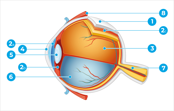
- 1 - Sclera
- 2a - Iris
- 2b - Ciliary Body
- 2c - Choroid
- 3 - Retina
- 4 - Cornea
- 5 - Crystalline Lens
- 6 - Vitreous
- 7 - Optic nerve
- 8 - Superior rectus muscle
Sclera: This is the outer layer of the eyeball, which takes up 4/5 of the posterior eye. It is known as the ‘white of the eye’, which is a layer that surrounds the outer part of the eyeball. The utmost anterior chamber of this layer comprises the cornea (described further on in the chapter on transparency).
Uvea: This is an essentially vascular layer and its name is due to the fact that it is intersected by blood vessels. It is divided into three portions: iris, ciliary body, and choroid.
Iris: This is the coloured part of the eye. It is a round structure with a central opening called pupil. The iris is located between the cornea and the crystalline lens. It functions as a type of photo camera shutter: when it is exposed to a great amount of light, its aperture in the middle decreases while on the other hand, when it is exposed to a small amount of light, it dilates and increases in diameter. Its job is to control the amount of light that goes into the eye and it plays an essential role in the eyesight with a black yet entirely transparent appearance. Every image we see goes through it.
Ciliary Body: It is made up of the ciliary processes that produce the aqueous humour and the ciliary muscle, which contracts causing the crystalline lens to accommodate.
Choroid: This is a vascular layer located between the sclera and the retina with nutritional functions, where the latter is the main responsible for turning the posterior segment of the eye into a dark place, which is crucial for a good eyesight.
Retina: This is the internal layer surrounding the posterior portion of the eyeball and it has the most important role in terms of eyesight. It comprises a large number of light-sensitive cells known as photoreceptor cells: the cones (responsible for colourful and daytime vision) and rods (responsible for black and white and night-time vision).
Cornea: This is the prominent anterior part of the eyeball, which is protruding and visible. It is entirely transparent and forms the outer layer of the eyeball together with the sclera. The corneal curvature is not spherical: it is slightly more significant vertically than horizontally. This difference in terms of curvature may be observed in different directions originating in most types of astigmatism. The cornea is an extremely important element in the dioptre of the visual system, since due to its significant curve it is the main way the parallel rays from infinity converge and enter the retina.
Aqueous Humour: This is a liquid and transparent substance that fills the anterior chamber of the eye (between the cornea and the iris) or the anterior segment of the eye (between the cornea and the crystalline lens) produced by the ciliary body, which due to its internal pressure leads to the cornea protruding and causing tension in the eye. Aqueous humour is constantly renewed and drained by means of a trabecular meshwork (in the angle between the cornea and the iris) passing on to Schlemm's canal. When this drainage is hard due to the structure being obstructed, there will be excessive pressure in the eye: an important risk factor for glaucoma.
Crystalline Lens: The crystalline lens is a transparent, biconvex biological lens located right behind the iris, between the eye's anterior and posterior chambers. Its job is to allow for a clear vision in every distance. When you look close, the crystalline lens will increasingly converge and its refractive power (accommodation) will increase and when you look far, it will converge less and its dioptric power will decrease. Its capacity to accommodate will decrease with age becoming increasingly hard to see close objects (presbyopia).
Vitreous Body: Also known as Vitreous Humour. This is an entirely clear jelly-like substance that takes up the inner space of the posterior segment of the eyeball causing it to take the approximate shape of a sphere.
Optic nerve: This is a group of tubular-shaped nerve fibres leading to the images caught by the retina to the cerebral cortex.
Extra-ocular muscles: The extra-ocular muscles limit the eyeball's movements. These six muscles – superior rectus, inferior rectus, medial rectus, lateral rectus, superior oblique, and inferior oblique – make the eye turn in all directions and act in agreement with both eyes enabling them to remain parallel.
Sight physiology: The eye is one of nature’s most resourceful devices. You may understand the whole mechanism of sight better if you compare the eyeball to a photo camera: the cornea and the crystalline lens are the camera lens where the latter is able to focus on objects at every distance; the iris is the shutter; and the retina is the film. This way, the light rays enter the cornea and aqueous humour passing through the pupil, they reach the crystalline lens, which focuses on the image further behind or further ahead allowing for it to project onto the retina. The image that is formed on the retina is reversed like in a photo camera. The amplitude of the visual angle is almost 180 degrees. The retina conveys visual information through the optic nerve to the cerebral cortex. Then, the brain begins to analyse and interpret (visual perception) enabling us to recognise colours, movement, distance, etc.