
The first computer appeared in 1939, in the United States. There has been a considerable technological evolution ever since, which had an inevitable impact on several aspects of everyday life.
Computer screens are the most important part of a computer in terms of eye impact. Screens are in fact used on a daily basis for work, education, and leisure.
When you talk about viewing screens, you should also consider TV and computer games. Television has a different kind of impact from computers, since in this case we should not only consider long-sightedness, which originates in lower vision concentration and mostly in convergence insufficiency.
For professional or private reasons, today there are in fact a large number of people spending several hours per day in front of a computer screen.
There have been several studies to check whether a viewing screen was responsible for many eye complications. The emission of dangerous electromagnetic radiation and the cause for cataracts have not been confirmed. In fact, the ultraviolet radiation produced by computers seems to be lower than that produced by a fluorescent lamp.
Another problem is eye strain. There, you have many associated eye disorders and eye discomfort.
When faced with an individual showing symptoms after working in front of a computer screen, it will be harder to differentiate between the causes of these symptoms and the “favourable” factors.
Thus, we may think that the symptoms resulting from working in front of a computer screen will be the result of eye disorders or an unfavourable eye environment, or even a combination of the two.
This way, we can consider three main causes/factors for these symptoms to appear when working in front of a computer screen.
The first factor is the group of eye causes. We can subdivide this group into a) uncorrected or badly corrected refractive errors, accommodative dysfunction, convergence insufficiency, and eye movement disorders; b) eye pathologies, such as allergy or dry eye syndrome; c) using contact lenses.
Environmental or ergonomic factors are the second factor. We can also subdivide them into a) conditions in the workplace, namely lighting, computer screen resolution, computer screen glare, and reflections. It should also be pointed out that the computer screen should not reflect artificial lights or light coming from a window, for which reason a computer screen should never be positioned in front of a window; b) we must also consider the overall conditions in the workplace, which may include the level of ventilation, heating, the impact of air conditioning, stress, and how work and breaks are organised.
The third factor with potential implications on the symptoms appearing after working in front of a computer screen is the overall health of the individual, which may include not only physical or emotional disorders but also skin pathologies like rosacea and seborrhoea.
Today, there is even a syndrome called Computer Vision Syndrome, which is characterised by eye strain associated with the long-term use of a computer. It is characterised by irritated eyes, red eyes, itch, dry eyes, fatigue, light sensitivity, the feeling of heavy eyelids, and difficulty in focusing on images.
Computer screen users ought to permanently obtain clear images, so this will easily result in the eye muscles straining. And the extra-ocular muscles are histologically striated muscle tissue, which is exactly the same as the muscle tissue on the legs. Hence we have fatigue when muscle (or eye) strain occurs for long periods of time.
- ophthalmic pathology (myopia, hypermetropia, and reduced short-sightedness)
- inappropriate environmental conditions (incorrect lighting and sitting posture and user-screen position)
- wrong use of screen (no breaks)
- By correctly positioning your computer 50-60cm (19-23in) away from your eyes. The upper line of the screen ought to be 2cm (0.7in) below the horizontal line of your eyes. Adjust the computer about eight degrees up. A good posture is fundamental for reducing muscle and eye strain.
- Check your posture. Your spine and neck ought to be straight in order to avoid the neck muscles from tensing up, since they are related to the extra-ocular muscles. Your hands ought to be higher than your elbows.
- If you are using documents, you ought to have them fixed laterally on a support by the computer as to avoid making excessive movements with your head.
- Good lighting in the area. It is fundamental that you minimise any computer screen reflections, be it from a window or any lamps.
- It is important to take breaks. Overall, a 5-10 minute break is recommended every two hours. In order to prevent eye strain, close your eyes for a few seconds and focus on distant objects for a few seconds.
The sun has benefits and harmful effects. Like in most things and attitudes, you need to determine the right dose. There are many benefits, which we know to be necessary. The sun is an anti-depressant and even anti-stress. It regulates our circadian rhythm, it heals some skin diseases, and since it stimulates the synthesis of vitamin D, it makes it easier to absorb phosphorus and calcium. It is also a positive influence on humour... and even a small ray of sunshine. However, sun rays ought to be enjoyed in moderate amounts, since they can be harmful if they are not taken in proper doses. If you absorb an excessive amount of sun rays, particularly at times when the sun is more intense, it can be damaging not only for your skin but also, and above all, on the eyes.
Due to its ‘visible light’, in addition to an unpleasant dazzling feeling, you are also left with a burning and tearing sensation.
The sun also produces rays with ultraviolet and infra-red wavelength.
Infra-red rays heat up the tissues drying the ocular surface and reducing the tear film, thus minimising its lubricating protection, which is necessary for the eyeball.
Ultraviolet rays go through the eye’s different optical media – namely the cornea and crystalline lens – and reach the retina. Excessive sun exposure will inevitably lead to early ageing of the retina's cells, which are essential to your eyesight. And if you look directly at the sun you can even go blind. Watch out when looking at sun eclipses!
The number of ultraviolet rays reaching us depends on the temperature, the distance between the sun and the earth, and the atmosphere: ozone layer, cloud density, and even pollution. It is most dangerous from noon to 4pm. But some ultraviolet rays – namely UVA – penetrate deep into the skin and are also present early in the day and at the end of the day.
Another aspect is snow, where about 80-90% of ultraviolet rays are reflected. Skiers beware! And sunglasses are not enough! You need to wear dark glasses with a high percentage of dark (more information later on).
You choose the density of your dark glasses based on the planned activity and particularly your environment. As is the case with sun protection, each level of protection is also characterised by an index. And naturally, we are not all the same when facing the SUN.
In fact, from the age of 60, adults are particularly sensitive to light given that during opacity the crystalline lens increases the dissemination of these rays and stimulates dazzling. Children are mostly in danger because they do not have an ultraviolet ray filter like adults do. So, there is more reason for children to wear sunglasses than adults.
But how do you choose sunglasses among a whole variety of brands and models with different shapes and prices sold in the market?
Good sunglasses ought to act not only in the dazzling effect but also against infra-red and ultraviolet rays. So, you need to check whether they comply with the EC standard ensuring a 100% anti-UV filter and also control with the help of an optician that they will protect you sufficiently against light according to use.
In the case of infra-red rays, there is no legal standard required but some manufacturers indicate the capacity of absorption of those lenses. It is also important to ensure that they do not change the perception of contrast nor do they cause visual disturbance.
The lateral protection of sunglasses is usually neglected. The more intense the sunlight, the more you will need lateral protection, which you ought to have when using dark glasses in the snow.
On paper, and generally speaking, lighter eyes will be more sensitive to sunlight. But the truth is that higher or lower light-sensitivity will depend not only on your eye pigmentation but also on other factors, such as: muscle strain, stress, and the eye’s refractive errors.
European standards classify sunglasses into four categories according to the percentage of their shade:
- CATEGORY 4 – recommended for high mountains and glaciers. Forbidden in road traffic.
- CATEGORY 3 – ideal for boats and sports under intensive lighting conditions.
- CATEGORY 2 – driving, beach, and terraces.
- CATEGORY 1 – driving, weak environmental light.
- It is a complication of Diabetes Mellitus in the eyes. It is usually progressive.
- It is serious! It is potentially serious but there is a solution! However, it is still the main cause of blindness between the ages of 30 and 60.
- Long-term hyperglycaemia will lead to changes in the retina’s blood circulation. You can compare blood vessels to hoses. These hoses may be blocked and have little holes in them where water begins to leak. This is exactly what happens with the retina's blood vessels. On one hand, a retinal vessel occlusion (leading to a reduced blood flow in the retinal vessels and subsequent ischemia) and blood and fat leak into the retina due to the barrier’s loss of function.
- Diabetic retinopathy is a disorder of multi-factorial causes. The risk factors that increase the chance of diabetic retinopathy are:
- age (it is rare before puberty).
- race (there is a higher incidence on the black race).
- genetics (four percent higher risk with a relative that has diabetic retinopathy).
- the duration of diabetes (having diabetes for more than 16 years will highly increase the risk of diabetic retinopathy. The longer the disease, the higher the risk of diabetic retinopathy).
- metabolic control (the greater the metabolic lack of control, the higher the risk of diabetic retinopathy).
- eye factors (patients with myopia have a lower risk of getting diabetic retinopathy).
- systemic factors (patients with anaemia, nephropathy, and autonomic neuropathy have a higher risk of getting diabetic retinopathy).
- several factors (smoking and pregnancy increase the risk of diabetic retinopathy appearing and developing).
- There are none at an early stage, which justifies the need for regular eye exams and monitoring in patients with diabetes “mellitus”.
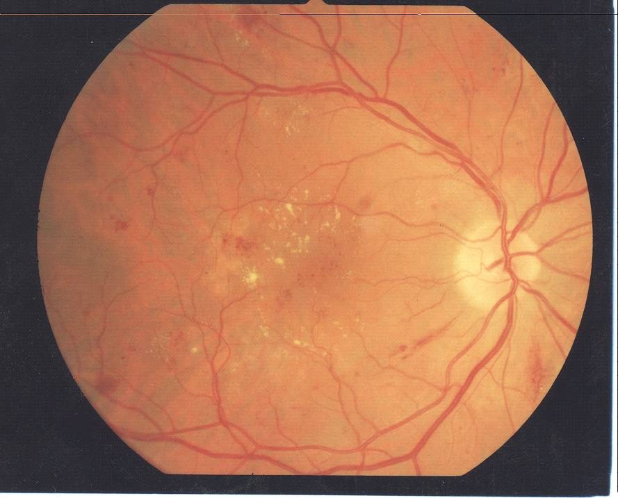
- There are three stages: background retinopathy (there is haemorrhaging in the retina, hard exudates, which are yellow lesions, and other changes), pre-proliferative retinopathy (extensive areas of ischemia in the retina), and proliferative retinopathy (abnormal vessels are present, which cause significant bleeding – neovessels. These abnormal vessels are created by the human body in order to try and fix the areas of ischemia in the retina.
- The most important treatment is panretinal photocoagulation together with a better metabolic diabetes control. In the case of haemovitreous (bleeding in the vitreous cavity) or preretinal membranes, you can have eye surgery called vitrectomy. More recently, in the case of a macular oedema that is resistant to the laser treatment or in the case of proliferative retinopathy, you may undergo an intravitreal injection of anti-angiogenic agents.
- It is fundamental and important for you to:
- Metabolic control! Metabolic control! Patients ought to try and get their blood sugar level to be regularly under 160 mg/dl. And also having a glycated haemoglobin test, which reflects metabolic control for the past three months.
- Regular exercise. Walk! Walk every day.
- Prevent and control some pathologies that influence diabetic retinopathy in a bad way, such as: anaemia, dyslipidemia, nephropathy...
- Avoid smoking.
- Regular eye tests, annually in mild cases and every three months in more severe cases.
- This is a serious eye condition and one of the main causes of blindness around the world. It is a disorder of the optic nerve, where the latter is affected and progressively and slowly destroyed as a consequence of increased intraocular pressure.
- There is a liquid inside the eye called aqueous humour. It is produced inside the eye and leaves the eye contributing to the eyeball’s shape and tone.
- The increase in intraocular pressure is caused when the aqueous humour becomes accumulated as a result of blockage to its draining or its increased production.
- When undiagnosed or untreated, it progressively and slowly destroys the optic nerve... evolving to blindness if left untreated! Increased intraocular pressure will make it difficult and block irrigation of the nerve fibres making up the optic nerve leading to its death.
- One in 100 forty-year-olds has some sort of glaucoma and one in ten 70-year-olds has some sort of glaucoma.
- It can appear at any age, but it occurs more frequently after the age of 30. The risk of having glaucoma increases considerably when there is a history of this disease in the family.
- The most common type is chronic open-angle glaucoma, which occurs in 90% of the cases. It is a slow and progressive type, which does not usually have symptoms. Increased intraocular pressure is mostly the result of increased difficulty in draining the eye, but it is also an increase in the production of aqueous humour.
- Acute closed-angle glaucoma is rare, serious, and has symptoms like painful eyes, red eyes, reduced sight, and vomiting. It is an eye emergency.
- Congenital glaucoma is rare.
- Secondary glaucoma is a result of systemic or eye disorders and medication.
- There are usually no symptoms. It progresses slowly and may take months or years to affect the edges of your vision (peripheral vision). Usually, at this stage, there is already a severe and irreversible lesion of the optic nerve.
- No! There is only ocular hypertension – without a glaucoma – in the case of increased intraocular pressure with normal optic nerve and vision.
- Besides, a glaucoma may also occur with a normal ocular tension.
- Nobody knows exactly. There are many factors but decreased vascular irrigation is one of the most important factors.
- It occurs when intraocular tension suddenly increases. There you have symptoms like painful eyes, reduced sight, halos around lights, red eyes, nausea, and vomiting.
Having/Being:
- over the age of 45.
- a family history.
- diabetes.
- systemic arterial hypertension and systemic arterial hypotension.
- taken medication (corticosteroid agents).
- black.
- It varies! It is higher in the morning. But these physiological variations alone do not cause eye lesions.
- Yes. It is a bilateral disease.
- It mostly depends on whether it is detected early on.
- There is no cure for glaucoma. But there is a treatment that aims at stopping the disease from progressing. You can take medication like: eye drops to decrease intraocular tension, pills to decrease eye tension, laser treatment, and surgery.
- Examine the fundus of the eye.
- Measure intraocular tension.
- Changes in vision.
- An average decrease in the thickness of nerve fibres.
- It is important to detect and diagnose a glaucoma as early as possible so that you may begin treatment.
- It is a silent and serious disease... for which reason you ought to have your eyes tested regularly.
- Changes in vision.
- If you have any questions, ask your ophthalmologist.
- These are allergic eye reactions, that is, they are the response of the immune system (the body’s system of defence) to a certain substance called allergen.
- It is a substance capable of producing allergic reactions.
- Pollen (trees, flowers, and some herbs, which release pollen into the air).
- Mould (mould spores in leaves and hay, which are released into the air).
- Animal hair (namely cat hair).
- Dust mites, pollution, cosmetic products, and make-up.
- Seafood (shrimp and lobster).
- Allergic conjunctivitis and allergic eyelid reactions.
- It is an inflamed conjunctiva. The conjunctiva is a thin and transparent membrane covering the white of the eye (sclera) and the internal surface of the eyelids.
- Yes. If the conjunctiva is exposed to an allergen it will lead to irritated blood vessels, which will subsequently dilate originating in a red eye.
- Yes. It may solely manifest itself through allergic conjunctivitis when people come into contact with dangerous substances blown in the wind like fungal spores, dust, and pet hair.
- Pruritus (itching).
- Watering eyes (tears).
- Burn.
- Eyelids (oedema, erythema, and skin peeling).
- Conjunctival hyperaemia (red eyes).
- Chemosis (swollen conjunctiva with a jelly-like appearance).
- An oedema and skin peeling around the eyelids.
- Seasonal or perennial allergic conjunctivitis (hay fever). In seasonal conjunctivitis, the most common allergen is grass or weed pollen. In perennial conjunctivitis, the most common allergens are dust mites and animal hair.
- Vernal Keratoconjunctivitis – this is rare but potentially serious due to reaching the cornea. Often, it is not possible to establish a prevailing allergen. It usually lasts five to ten years.
- Atopic Keratoconjunctivitis – it may lead to blindness due to reaching the cornea. It may last decades.
- Papillary Conjunctivitis – it is usually the result of being intolerant to contact lenses and/or products used to disinfect contact lenses.
- Contact or toxic conjunctivitis – an allergic reaction to some drugs (pain killers, Atropine, Chloramphenicol, Gentamicin, etc.) and preservatives.
- Anyone is prone to allergic eye reactions. However, people suffering from asthma, allergic rhinitis, or skin allergies are at higher risk of contracting eye allergies.
- Yes. It is usually self-limiting lasting five to ten years. Atopic keratoconjunctivitis is an exception, which may last decades.
- Yes, for two reasons. The first reason is that scratching your eyes will stimulate the emergence of chemical substances, which will then stimulate itchiness (this is a vicious cycle). The second reason is that in the case of people with atopic keratoconjunctivitis, scratching your eyes will lead to a corneal disease called keratoconus.
- Keep environments free from dust.
- Wash clothes you have kept away for a long time before wearing them.
- Avoid dusty and smoky environments with strong odours.
- Get rid of fabric that will accumulate dust like mats, carpets, curtains, and soft toys.
- Keep air conditioning filter clean.
- Sleep in synthetic bedding and avoid wool duvets, quilts, fluffy blankets, and flannel bed sheets.
- Avoid sofa cushions and use plastic covers at all times.
- Avoid decorative objects that may accumulate dust.
- Avoid plants, flowers, and pets with hair inside the house.
- Dust frequently with a vacuum cleaner or damp cloth at all times.
- Avoid contact with pets.
- Avoid walking through fields, particularly in the spring and autumn, and particularly on windy days.
- Try to keep your windows closed in the spring and autumn.
- Vacuum your mattress every week.
- Keep objects that may accumulate dust like soft toys and books inside cupboards and drawers.
- Replace your broom with a vacuum cleaner.
- Avoid taking medication that may have caused you allergic reactions in the past.
- Cover pillows and cushions with waterproof covers.
- Limit the use of cosmetic products and make-up.
- Avoid wearing contact lenses.
- Avoid going into swimming pools without goggles and a seal.
- Avoid allergenic food (bananas, strawberries, kiwi, and seafood).
- When strolling outdoors protect your eyes by wearing sunglasses.
- Avoid rubbing or scratching your eyes.
- Quickly leave the place where the symptoms appeared.
- Wash your eyes with a physiological solution.
- Do not rub your eyes.
- Apply cold pads on your closed eyes.
- Make an appointment with an ophthalmologist to begin your treatment.
- The only cure is by avoiding allergens (do not contact these allergens anymore).
- You can apply a topical (eye drops and eye ointment) or systemic treatment to relieve the symptoms.
- This is a clinical condition where there is a quantitative and/or qualitative pre-corneal tear film deficiency, which is multi-factorial and results in the cornea not being properly hydrated.
- It is the loss or reduction of the normal capacity of the eyes to produce tears... a balance is necessary between the production of tears and their excretion either by draining or evaporation.
- When you lose that balance the tear film begins to rupture producing dry areas on the cornea and conjunctiva leading to lesions and subsequent symptoms.
- When it is not diagnosed and/or treated properly it may lead to severe lesions on the ocular surface, and in more severe cases, to blindness.
- In the main and accessory lacrimal glands.
- The main lacrimal glands, which are located in the eye socket and eyelids, produce reflex tears in response to a stimulus (irritation, emotion). However, these glands do not contribute to the eyeball's hydration paradoxically leading to people suffering from dry eyes complaining that they cry a lot.
- The accessory lacrimal glands are good for hydrating and nourishing the cornea making it easier to see clearly and removing foreign matter and micro-organisms. These are basal tears.
- Tears are constantly produced so that the ocular surface is hydrated. This production may increase, as mentioned before, as a consequence of responding to an external stimulus like trauma, emotions, allergies, or air pollution.
- Normally, blinking will spread the tears evenly through the ocular surface as a thin layer – the tear film –, which will break after a few seconds and you will need to blink again.
- We blink an average of 15 times per minute.
- Blinking will lead to tears covering the corneal surface hydrating and protecting it and nourishing your eyes. It also allows for a clear vision with no distortions.
- Burn.
- Irritation.
- The feeling of having a foreign matter or sand in your eyes.
- Excessive watering eyes (confusing symptom!).
- Photophobia (extreme sensitivity to light).
- Blurred vision.
- Red eyes.
- Discomfort after reading, watching TV, or working on a computer.
- Mucous secretion in your eyes.
- Tears are secreted by the main and accessory lacrimal glands. They are drained at the bottom of a conjunctival sac and then eliminated in the nasal cavities through excretory ducts.
- This is an important structure that ensures the physical integrity of the cornea and conjunctiva.
- Lipid layer (the most anterior layer) – it is secreted by the Meibomian glands. Its job is to delay the evaporation of the aqueous layer (middle layer) thus stabilising the tear film.
- Aqueous layer (middle layer) – it occupies almost all of the tear film’s thickness (98% of thickness). It also carries oxygen to the corneal epithelium; it contains lysozyme and beta-lysine (bacterial function) and immuno-globulins (particularly IgA, which stops bacteria from adhering to the corneal surface). This layer also plays an important role in maintaining image clarity.
- Mucin layer (intimately connected to the corneal epithelium and bulbar conjunctiva) – the mucin layer is a layer that adheres to the surface of the corneal epithelium leading to tears being evenly distributed on the corneal surface in-between blinking.
- It is certainly one of the most important causes of eye discomfort in the elderly.
- It is estimated that 15-40% of the population above the age of 65 is affected by this pathology.
- There may be systemic diseases, but most people do not show these pathologies.
- Activities that require greater concentration, such as reading, using computers, driving, and watching TV, that is, whenever you require greater concentration resulting in a decrease in the number of times you blink.
- Watching TV above your eye level will cause your eyelids to open more leading to a higher evaporation of tears.
- Environmental conditions, such as wind and air conditioning, which will lead to a higher evaporation of tears.
- Dry eyes occur more often in autumn and winter due to low air humidity.
- Menopausal women due to hormonal changes.
- Age (fewer tears are produced as we grow older. For instance, at the age of 65, you produce less 60% of tears than at the age of 18).
- Systemic causes: Rheumatoid arthritis, systemic lupus erythematosus, scleroderma, polyarteritis nodosa, sarcoidosis, Parkinson's disease, Diabetes Mellitus, and thyroid disease.
- Eyelid and corneal epithelium abnormalities.
- Drugs that decrease the production of tears (antihistamines, tranquillisers, decongestants, anti-depressants, oral contraceptives, pain killers, and beta-blockers).
- Postoperative stage for refractive surgery.
- Contact lenses (they may lead to dryer eyes or worsen a pre-existing condition).
- Remaining at higher altitudes.
- Smoking. Air pollution.
- Dry and windy environments.
- Chemical burns.
- A completely examination of the patient and specific tests are carried out (break-up time test, Schirmer's test, and Rose Bengal). That is, observation and therapeutic orientation by an ophthalmologist.
- It is palliative! Systemic pathologies are observed and predisposing factors are corrected. Artificial tears, preferably with no preservatives. In the form of eye drops or gel in more severe cases. Punctual occlusion (in more severe cases).
- They are called “MUSCAE VOLITANTES” (flying flies) in Latin. This is also called MYODESOPSIA, which comes from the Greek “MYODES” (similar to flies) and “OPSIS” (sight).
- This is an eye defect (symptom) that manifests itself in our vision as spots, filaments, or dots in our visual angle.
- They are vitreous humour opacities (condensation), the transparent jelly filling the eyeball, which are hit by a stream of light and project a shadow onto the retina as the eye moves.
- They may have different shapes, spots, lines, cobwebs, flies, clouds, filaments, circles...
- They are caused by physiological changes in the vitreous as a result of age or eye diseases. The vitreous gel begins to shrink, it peels away from the posterior wall of the eyeball (retina) causing a posterior vitreous detachment.
- It is a jelly-like structure that fills the entire posterior cavity of the eyeball.
- Its fundamental characteristic is transparency.
- Vitreous gel is in contact with the retina's entire surface.
- Vitreous gel consists of 99% of water and 1% of solid elements like collagen fibres, hyaluronic acid, and proteoglycans.
- They are shadows projected onto the retina.
- They are uncomfortable and annoying right when they appear although people may adapt in time.
- It depends on where they are located, they may interfere with reading.
- They are more noticeable when looking at a plain background like a white wall or blue sky.
- They usually appear at the age of 40 and above, or earlier in the case of people with myopia.
- With age, the vitreous fibrils degenerate originating in a vitreous contraction.
- They appear in the form of dark spots like ‘flies’ or ‘cobwebs’ when the eyes move.
- The most accepted hypothesis is that this change in the structure of the vitreous gel is the result of changes in the macromolecules within.
- Myopia.
- After cataract surgery.
- After YAG Laser.
- After eye inflammation (posterior uveitis).
- After eye trauma.
- Do not follow these opacities with your eyes because it may lead to traction on the retina and originate in a torn retina.
- Avoid looking at light and plain surfaces.
- Be patient.
- They may be serious when associated with a torn retina, since if left undetected and untreated it may lead to a retinal detachment.
- There is no efficient or safe treatment for myodesopsia.
- In the case of causal, inflammatory, and vascular pathologies, the treatment is etiologic.
- They rarely disappear physically.
- In the case of large floaters interfering with your eyesight, you may be treated with a YAG Laser.
- An ophthalmologist observing the fundus of the eye, namely around the retina, and laser photocoagulation in the case of a torn retina.
- Or Photopsia!
- They occur when the vitreous gel pulls on the retina after degeneration.
- It is a mechanic stimulus exercised directly on the retina.
- In the case of sudden flashing lights, you should make an appointment with an ophthalmologist to check for a torn retina.
- When new floaters appear.
- When flashing lights appear.
- When loss of the peripheral vision occurs.
Content taken from the website www.antonioramalho.com , with the author's knowledge and consent.