The ears are paired organs located on either side of the head and play a fundamental role in hearing and balance1.
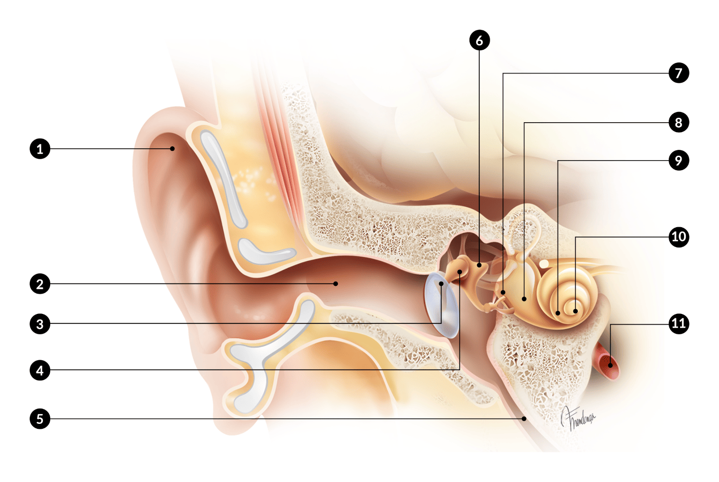
The hearing system is divided into the outer ear, middle ear and inner ear2 (Fig. 1) and each of these parts plays an important role in sound processing and balance.
The outer ear is made up of the pinna (commonly known as the ear) and the External Auditory Canal up to the tympanic membrane.
The middle ear is an airy cavity behind the tympanic membrane, contains the ossicles and communicates with the back of the nose via narrow canals, the Eustachian tubes.
The inner ear is located in the temporal bone, deep in the skull1,2.
https://otosurgeryatlas.stanford.edu/otologic-surgery-atlas/surgical-anatomy-of-the-ear/
HOW DOES HEARING WORK?
The outer ear receives and conducts sound to the middle ear, which amplifies it and conducts it to the inner ear. In the inner ear, sound is transformed into electrical signals that are sent to the brain via the auditory nerves. The inner ear also plays a role in the balance of the body1,3,4.
OUTER EAR
The pinna (ear) is the part of the ear that can be seen externally on either side of the head. It has the shape of a cone with an irregular surface, inclined at 30°, which makes it easier to pick up sound coming from the front. The various features of the pinna play a role in detecting the location of the sound source1,3.
Sound is conducted through the ear canal, ending at the tympanic membrane (or eardrum), a thin elastic membrane that vibrates and borders the middle ear. This canal is covered with skin and has cerumen-producing glands1,3,4.
MIDDLE EAR
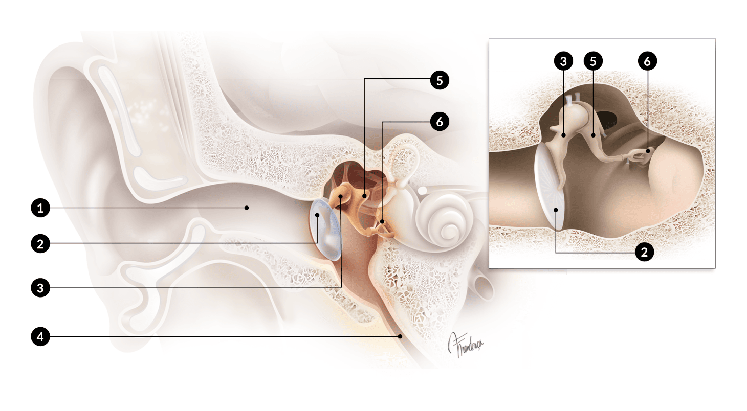
The middle ear is an airy cavity located behind the tympanic membrane. It communicates with the back of the nose through a narrow canal named the Eustachian tube, which allows to normalise pressures and drain mucus from the middle ear. Equal air pressure on both sides of themembrane is important for good hearing4.
This cavity houses small bones - the malleus, incus and stapes - which act as amplifying levers for the sound transmitted to them by the eardrum, amplifying it in the direction of the inner ear1,3,4 (Fig. 2).
https://otosurgeryatlas.stanford.edu/otologic-surgery-atlas/surgical-anatomy-of-the-ear/
INNER EAR
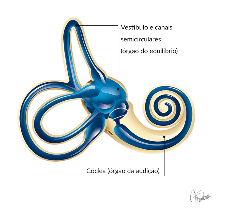
The inner ear is located in the temporal bone, in the depths of the skull. It groups together two sensory organs - the vestibule (which includes the semicircular canals), which is the organ of balance, and the cochlea or snail, which is the organ of hearing1,4 (Fig. 3).
https://otosurgeryatlas.stanford.edu/otologic-surgery-atlas/surgical-anatomy-of-the-ear/
The structures of the inner ear are filled with fluids that circulate in a system of small canals and saccules - the utricle and saccule and the vestibular and tympanic ramps. The vestibule and the cochlea contain microscopic cells and structures that have sensors that are stimulated by fluids and send balance and hearing signals to the brain via the vestibulocochlear nerve.
1. Dr. Brian Yeom, Waikato Clinical Campus https://www.entinfo.nz/anatomy-of-the-ear/ consultado em 16-11-23
2. Dr. Jackler and Ms. Gralapp https://otosurgeryatlas.stanford.edu/otologic-surgery-atlas/surgical-anatomy-of-the-ear/ consultado em 16-11-23
3. Auditory Anatomy and Physiology, Trigueiros N., Subtil J., in Otoneurologia, 29-38, 1ª Edição 2023, Ed. Círculo Médico
4. https://www.mayoclinic.org/diseases-conditions/hearing-loss/in-depth/ear-infections/art-20546801 consultado em 16-11-23